Upper GI Endoscopy(EGD)
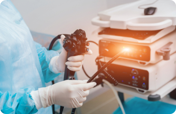
Upper GI endoscopy is also known as esophagogastroduodenoscopy and EGD. EGD is used to visually examine your entire esophagus, stomach and duodenum (the first portion of small intestine) for abnormalities. It is generally considered the procedure of choice for evaluating the lining of the mentioned organs. During the exam, an endoscope — a long, flexible tube about the thickness of an adult finger — is inserted into your mouth. A tiny video camera at its tip allows your doctor to view the inside of your upper GI tract.
In some cases during EGD, if a polyp or abnormal tissue is found, your doctor may remove it at that time. Alternatively, a tissue sample (biopsy) of the polyp may be taken for lab analysis to determine whether subsequent surgical removal of the tissue is needed. Contact us for more information about Upper GI Endoscopy(EGD) in Beverly Hills

How do you prepare?
For the EGD procedure to be accurate, your stomach must be empty. Thus no food or drinks should be consumed for at least eight hours prior to the procedure. Dr. Davidson will review your past medical history and medications and will notify you which medications should be held on the morning of the exam.
If you have diabetes or take blood thinners, including aspirin or other pain relievers, your preparation for EGD may be slightly different. Notify your doctor of either of these factors at least seven days ahead of the test, to see if you need additional instructions.
How is it done?
Upper GI endoscopy can be relatively painless when performed by an experienced practitioner. You will receive a sedative medication administered intravenously to minimize any discomfort.
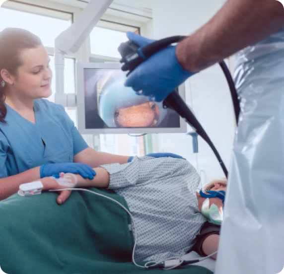
During the exam you’ll likely lie on your left side. Dr. Davidson inserts the endoscope in your mouth. The endoscope contains a fiber-optic light and a channel that allows your doctor to pump air into your stomach, inflating it to get a better view of the interior lining.
The endoscope also contains a tiny video camera at its tip. The camera transmits images to an external monitor so that your doctor can look closely at the inside of your body. Your doctor can insert instruments through the scope’s channel to remove polyps, take tissue samples, inject solutions or destroy (cauterize) tissues.
If a polyp or abnormal tissue is found, your doctor may choose to remove it with a snare or destroy it with cautery. Or he or she might take a biopsy or advise surgical removal, depending on the size of the mass.
An upper GI endoscopy exam usually takes less than 20 minutes.

After the procedure
After the exam is over, it takes less than an hour to recover from the sedative. You’ll need someone to take you home because it can take up to a day for the full effects of the sedative to wear off. Rest and don’t drive and do not sign any legal documents for the remainder of the day.
You may feel bloated or pass gas for a few hours after the exam. You should feel better as you pass the gas. Walking may lessen your discomfort. If you have persistent pain after the procedure, tell your doctor.
These signs and symptoms may result from bleeding when a biopsy is taken or, rarely, from perforation of the colon wall. Although they’re rare, be alert for these signs and symptoms, as they can indicate the need for medical attention.
Results
Frequency of follow-up exams depends on the findings as well as the quality of the exam performed and should be discussed with your doctor. If an ulcer, polyp or abnormal tissue was found during your endoscopy that couldn’t be removed, your doctor may recommend subsequent procedures.

Negative test results. You will be reassured. If further testing or treatment is indicated, Dr. Davidson will arrange for it.

