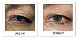
A pterygium (teh-RIJ-ee-um) is an elevated, wedged-shaped, clear to milky color growth of the scleral (the white part of eye) that invades the cornea. Pterygia are benign (non-cancerous) growths, but contain blood vessels and form scar tissue on the eye. Because a pterygium resembles tissue or film growing over the eye, a person who has one may become concerned about personal appearance. Risk factors for developing pterygium include prolonged exposure to ultraviolet light from the sun may play a role in the formation of pterygia.
Some people with pterygia do not experience symptoms or require treatment of pterygium. However pterygia may become red and swollen on occasion, and some may become large or thick. This may cause concern about appearance or create a feeling of having a foreign body in the eye.
Treatment depends on the pterygium’s size and the symptoms it causes. If a pterygium is small but becomes inflamed, your eye doctor may prescribe lubricants or possibly a mild steroid eye drop to reduce swelling and redness. In some cases, surgical removal of the pterygium is necessary.
The pterygium may be removed in an operating room setting and under a microscope. A number of surgical techniques are currently used to remove pterygia, and it is up to your eye doctor to determine the best procedure for you.
After use of local or some other type of anesthesia, the pterygium is surgically removed. After the procedure, which usually lasts no longer than half an hour, you likely will need to wear an eye patch for protection for a day or two. You should be able to return to work or normal activities the next day. To prevent regrowth after the pterygium is surgically removed, your eye surgeon may suture or glue a piece of surface eye tissue or conjuctiva onto the affected area. This method, called autologous conjunctival autografting, has been shown to safely and effectively reduce the risk of pterygium recurrence.

